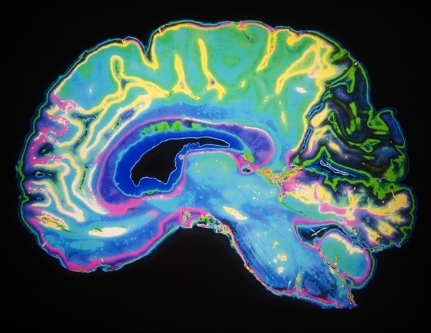
Researchers at Karolinska Institutet and Karolinska University Hospital have developed a groundbreaking microscopy method that enables detailed three-dimensional (3D) RNA analysis at cellular resolution in whole intact mouse brains. The new method, called TRISCO, has the potential to transform our understanding of brain function, both in normal conditions and in disease, according to the new study published in Science.
Despite great advances in RNA analysis, linking RNA data to its spatial context has long been a challenge, especially in intact 3D tissue volumes. The TRISCO method now makes it possible to perform three-dimensional RNA imaging of whole mouse brains without the need to slice the brain into thin sections, which was previously necessary.
This method is a powerful tool that can drive brain research forward. With TRISCO, we can study the complex anatomical structure of the brain in a way that was previously not possible.”
Per Uhlén, professor at the Department of Medical Biochemistry and Biophysics, Karolinska Institutet, and study’s last author
In the study, up to three different RNA molecules were analysed simultaneously. The next step for the researchers is to expand the number of RNA molecules that can be studied to around a hundred, using a technique called multiplex RNA analysis. This could provide even more detailed information about brain function and disease states.
The TRISCO approach opens up new possibilities to understand the complexity of the brain in depth, which in turn can lead to the development of new treatments for various brain diseases.
“We look forward to continuing our research and exploring the many possibilities offered by this new technique,” says Shigeaki Kanatani, a research specialist in Uhlén’s laboratory and the first author of the study.
Not only is TRISCO suitable for studying intact mouse brains, but the study demonstrates it can be used for larger brains, such as those of guinea pigs, and various tissues like kidney, heart, and lung. The study is a collaboration between Karolinska Institutet and Karolinska University Hospital.
“Our laboratory has several collaborations with clinically active researchers at Karolinska University Hospital. It is crucial for biomedical research that basic researchers and clinicians collaborate and understand each other,” says Per Uhlén.
The study has been funded by the Swedish Research Council, Swedish Brain Foundation and Swedish Cancer Society. Some of the co-authors are employed and own shares in the Danish company Gubra.
Source:
Journal reference:
Kanatani, S., et al. (2024). Whole-brain spatial transcriptional analysis at cellular resolution. Science. doi.org/10.1126/science.adn9947.
Source link : News-Medica

