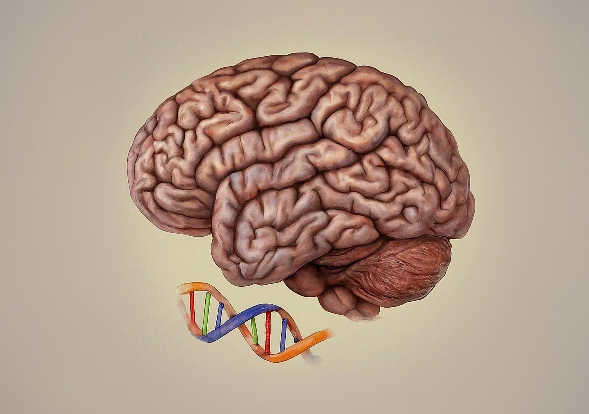
New insights into brain development: study maps chromatin changes linked to psychiatric disorders in prenatal and infant stages.
Study: Temporally distinct 3D multi-omic dynamics in the developing human brain. Image Credit: Shutterstock AI / Shutterstock.com
In a recent study published in Nature, researchers investigate chromatin conformational and epigenomic reorganization during the development of the prefrontal cortex (PFC) and hippocampus (HPC).
Early development of human brain
The human brain consists of hundreds of types of cells with diverse morphological, molecular, functional, and anatomical features. Most cortical neurons form during the first and second trimesters of pregnancy; however, the distinct molecular signatures of these cells emerge between the third trimester and adolescence.
Recent studies have reported chromatin remodeling in early postnatal development. Nevertheless, the trajectories of DNA methylation and chromatin conformational changes in prenatal brain tissues have not been characterized at single-cell resolution.
Investigating PFC and HPC development
In the current study, researchers investigate the dynamics of human PFC and HPC development using single-nucleus methyl-chromatin conformation capture (3C) sequencing (snm3C-seq3). About 29,700 snm3C-seq3 profiles were generated from adult and developing human frontal cortex samples, whereas over 23,300 snm3C-seq3 profiles were isolated from HPC samples.
The researchers quantified 3C information, as well as non-cytosine-guanine (non-CG) and CG methylation data, in individual cells at 100 kilobase (kb) resolution. A total of 139 cell populations were identified across developmental stages. Excitatory neurons exhibited distinct epigenomic types in the HPC and PFC, whereas non-neuronal and inhibitory neurons were shared between the two regions.
Non-CG methylation starts earlier in HPC inhibitory and excitatory neurons than in PFC neurons, with significant non-CG methylation observed in the HPC by the 39th gestational week (GW). In contrast, PFC neurons do not exhibit comparable levels of non-CG methylation until infancy.
Chromatin interactions and validation
Single-cell 3C profiles were subsequently clustered by the distance distribution between interacting loci. To this end, neuronal cell types were strongly enriched in clusters dominated by short-range interactions, whereas non-neural and glial cells were enriched in clusters dominated by long-range interactions.
A public and bulk Hi-C dataset of primary human tissues was also analyzed. This analysis indicated that chromatin conformation profiles from all tissues had the LE signature, thus suggesting that sex (SE) chromatin was specific to neuronal cells.
Chromatin tracing and ribonucleic acid (RNA) multiplexed error-robust fluorescence in situ hybridization (MERFISH) were also performed to validate the neuronal-specific SE chromatin conformation in newly differentiated neurons in mid-gestation (GW 23).
Chromosome 14 was imaged at 250 kb resolution by labeling 354 genomic loci, allowing the reconstruction of conformation of 46,023 homologs of the chromosome in 24,099 cells across the choroid plexus, fimbria, and HPC.
RNA MERFISH was performed on the same tissue sections. In neuronal cell types, genomic regions separated by short distances of up to five Mb had a compact spatial distance, thus indicating high interaction frequency.
Distal genomic regions exhibited larger physical distances, thus suggesting low interaction frequency. Non-neuronal cells and progenitors exhibited opposite patterns, as loci with short genomic distances had larger physical distances, whereas distal genomic loci had lower spatial distances. Imaging results also validated SE conformation emergence during the differentiation of radial glia (RG) to excitatory neurons.
Developmental changes in chromatin structure
Chromatin compartmentalization strength scores inversely correlated with the short-to-long-range interaction ratio. As a result, cell types with the LE conformation were associated with stronger compartment strengths. Compartment strength was also developmentally regulated, with a loss of strength observed between middle and late gestation, followed by a gain during further development.
An analysis of variance-based approach was utilized to identify differential chromatin loops across cell-type differentiation trajectories. Developmentally gained chromatin loops significantly outnumbered developmentally lost loops in all neuronal trajectories. In astrocytes, which exhibit the LE conformation, similar numbers of loops were gained and lost during differentiation.
Neuropsychiatric disorder heritability
Over 2.5 million differentially methylated regions (DMRs) were identified across developmental stages and cell types. Transcription factor (TF)-binding motif analysis revealed that the sequential action of lineage-specific and activity-dependent TFs shapes the regulatory landscape of inhibitory and excitatory neurons.
Regulatory elements that activate in mid-gestation become enriched in binding motifs of lineage-specific TFs. After lineage specification, the binding motifs of activity-dependent TFs become enriched in regulatory elements activated in late gestation and infant stages.
The researchers also quantified the polygenic heritability enrichment of annotations defined by chromatin loops or DMRs for each cell type. Loop-connected DMRs exhibited significantly greater heritability enrichment than all DMRs, thereby supporting the use of loops to locate causal variants.
A total of 190 fine-mapped putative schizophrenia causal loci were overlapped to DMRs and loop-connected DMRs. A robust correlation was observed between the odds ratio of overlapping with a putative causal variant and heritability enrichment across cell types.
The developmental dynamics of the enrichment of neuropsychiatric disorder heritability in neuronal populations was also assessed. This revealed similar patterns for DMRs and loop-connected DMRs over developmental trajectories; however, loop-connected DMRs had higher overall heritability enrichment.
The polygenic heritability enrichment for bipolar disorder and schizophrenia increased from neuro progenitors to early post-mitotic neurons and further to post-mitotic neurons in late gestation. Overall, there was a consistent developmental increase in heritability enrichment for these neuropsychiatric disorders between neuro progenitors and neurons in infants, which decreased in the adult brain.
Conclusions
The study findings highlight the dynamic transitions from progenitors to glial and neuronal populations from the second and third trimesters to the neonatal period. Remodeling of the neuronal chromatin conformation and methylome during perinatal development indicates that the human brain is susceptible to environmental and genetic perturbations.
The genetic risk of bipolar disorder and schizophrenia also appears to peak during the third trimester and infancy. Overall, the data provide multimodal resources to study gene regulatory dynamics in the developing brain and illustrate that single-cell multi-omics is a powerful tool to delineate neuropsychiatric risk loci.
Journal reference:
- Heffel, M. G., Zhou, J., Zhang, Y., et al. (2024). Temporally distinct 3D multi-omic dynamics in the developing human brain. Nature. doi:10.1038/s41586-024-08030-7
Source link : News-Medica

