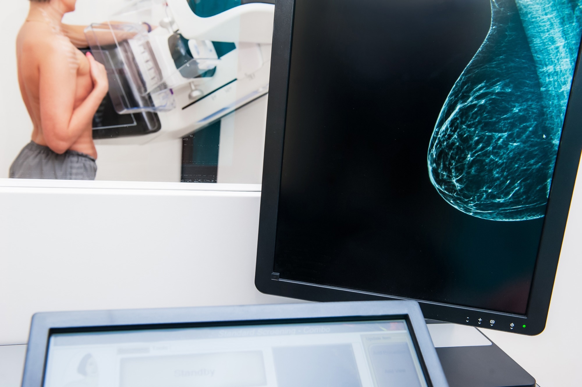
Using AI in mammogram screenings can help doctors identify breast cancer risks years before a diagnosis, opening doors to personalized, preventive treatments and more effective care.
Study: Artificial Intelligence Algorithm for Subclinical Breast Cancer Detection. Image Credit: Okrasiuk / Shutterstock.com
A recent study published in JAMA Network Open evaluates the efficacy of commercial artificial intelligence (AI) tools in detecting subclinical breast cancer from screening mammograms years before clinical diagnosis.
Integrating AI with mammography
In 2022, over 2.3 million women throughout the world were diagnosed with breast cancer, with over 670,000 deaths attributed to breast cancer that year. Within the United States, breast cancer is the most common type of cancer to affect women, with one in three new female cancers affecting breast tissues each year in this nation.
The United States Centers for Disease Control and Prevention (CDC) currently advises women 40 years of age and older to undergo a mammogram every two years to screen for the presence of breast cancer. Despite its widespread use, the accuracy of mammography is limited.
Recently, several AI algorithms have been approved to improve the accuracy of radiologist reports, which is achieved by marking suspicious regions and generating cancer scores to provide a more accurate diagnosis. In fact, some studies have suggested that these scores can predict future breast cancer risk before clinical features arise.
About the study
The current study included 116,495 women at nine breast centers in Norway who underwent three or more consecutive mammography screening rounds every two years. These mammograms were performed between September 13, 2004, and December 21, 2018.
All mammogram results were subjected to AI analysis using INSIGHT MMG, a commercially available AI algorithm. Importantly, INSIGHT MMG was not originally developed to estimate future cancer risk and has not been optimized for this task.
The algorithm provided a continuous variable, the cancer detection score, ranging from zero to 100. Higher scores reflected an increased risk of a positive mammogram.
Maximum AI scores and the absolute difference in scores were compared between the breasts of women screening positive for cancer, those negative for cancer, and those who developed cancer in the interval between mammograms.
Difference in scores
The study cohort comprised 1,265 and 116,495 women who screened positive and negative for breast cancer, respectively, as well as 342 women diagnosed with breast cancer during the interval period between mammograms. The average age at the third round for women screened positive for breast cancer was 58.5 years as compared to 57.4 and 56.4 years among women with interval cancer and screening-negative women, respectively.
The mean absolute differences (MADs) between the AI scores of both breasts were calculated. For cancer-free women, these differences were 9.9, 9.6, and 9.3 for each of the three rounds, respectively.
The MAD in AI scores of women who developed screening-detected cancer in the third round were 21.3, 30.7, and 79 in the first, second, and third rounds, respectively. Among those with interval cancer, MADs were 19.7, 21, and 34 in each round, respectively.
Scores were higher in breasts where cancer developed than in the other breast, with the increase present four to six years before the cancer was ultimately detected. The areas under the receiver operating characteristic curve (AUCs) discriminating between screening-positive and cancer-free women were 0.64, 0.73, and 0.97 at each round, respectively. AUCs increased from 0.66 to 0.78 for interval cancers at each round.
AUCs for the absolute differences in scores for screening-positive women were 0.63, 0.72, and 0.96 at each round among women detected by screening to have cancer. In contrast, interval cancer showed corresponding values of 0.64, 0.65, and 0.77 for each round. When all breast cancers were considered, AUCs increased from 0.64 to 0.93 between all rounds.
Interval cancers appear to develop faster and are more likely to be radiographically hidden in mammograms than those detected by screening rather than being missed by the radiologist.
Considering the top 1% of examination-level AI scores as positive for cancer and the remaining 99% as negative, the absolute score threshold was 91.3. At this threshold, 4.5%, 8.6%, and 53% of cancers would have positive AI scores across the three rounds, respectively. False-positive scores would arise for 0.7% of women in each study round.
Conclusions
The study findings indicate the potential to use AI mammogram scores to estimate breast cancer risk up to six years before diagnosis. The mean absolute AI scores were higher for breasts where cancer was developing than for the other breast, which was reflected in higher MAD scores.
Based on MAD scores, INSIGHT MMG accurately discriminated between women at an increased risk of future cancer as compared to cancer-free women. Using AI, women identified to be at a high risk of developing breast could then receive additional screening and other personalized interventions to prevent breast cancer.
The increasing difference in AI scores by time and between the breasts could be used by interpreting radiologists to indicate elevated risk of developing breast cancer.”
Source link : News-Medica

