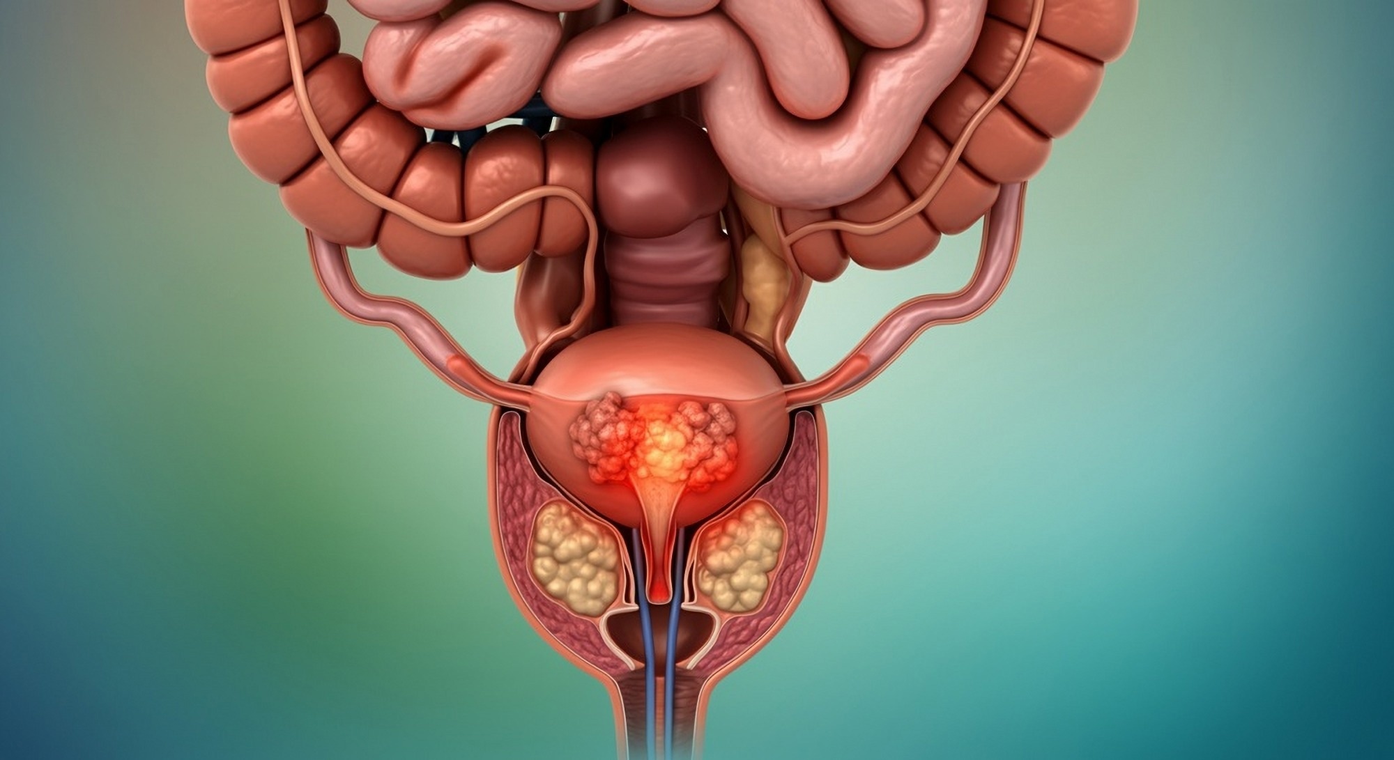
AI-driven MRI analysis offers new insights into prostate cancer prognosis, accurately predicting metastasis risk and treatment outcomes for improved patient care.
Study: AI-derived Tumor Volume from Multiparametric MRI and Outcomes in Localized Prostate Cancer. Image Credit: Shutterstock AI / Shutterstock.com
In a recent study published in Radiology, researchers determine whether measuring tumor volume inside the prostate using artificial intelligence (AI)-based magnetic resonance imaging (MRI) data could predict outcomes, including metastasis risk, for prostate cancer patients who have been treated with radiology or surgery.
Advances in MRI transform prostate cancer detection and diagnosis
Multiparametric MRI combines multiple MRI techniques to create detailed images of internal anatomy. This imaging technology has transformed the management of prostate cancer by improving the detection of serious cases while minimizing the detection rate of insignificant diseases. MRI-guided biopsies also significantly improve the accuracy of cancer diagnoses.
Various prostate cancer features are observed on MRI, some of which include prostate imaging reporting and data system (PI-RADS) scores, lesion scores, and radiologic T stage, the latter of which indicates the extent to which the tumor has spread within the prostate. Analyzing these features can indicate the potential recurrence rates of prostate cancer; however, the assessment of these features varies across observers. Various tumor grading systems are associated with differing accuracies, which further complicates diagnostic consistency.
The use of AI could enhance the clinical value of MRIs by providing consistent analysis of images. Recent studies on deep learning models have indicated accuracy levels comparable to that of experienced radiologists in outlining tumors within the prostate.
About the study
The current study aimed to determine whether calculations of tumor volumes using AI-based approaches can provide independent prognostic insights for prostate cancer patients who have previously undergone surgery or radiation therapy. These results were then compared to those obtained from standard MRI evaluations.
This retrospective study included prostate cancer patients who underwent MRI scans before undergoing either radical prostatectomy or radiation therapy. Patient data were gathered from medical records and consisted of clinical, pathological, and treatment information, including the classification of the tumor based on PI-RADS and National Comprehensive Cancer Network (NCCN) scores.
Biochemical failure is an increase in prostate-specific antigen (PSA) levels after treatments like radical prostatectomy or radiation therapy. For the current study, biochemical failure was defined as an increase in PSA concentrations by at least two ng/mL above the post-treatment lowest level for radiation therapy, and clinical progression or PSA increase of least 0.1 ng/mL in cases of radical prostatectomy.
Reference segmentations were manually created by a genitourinary radiation oncologist, who delineated prostate regions such as translational and peripheral zones and lesions with PI-RADS scores of three to five.
The AI model nnU-Net, a deep learning-based segmentation method, was trained to delineate prostate regions and tumors from different MRI sequences. The model was then validated using a subgroup of images from patients who received radiation therapy before being tested on images from both radiation therapy and radical prostatectomy groups. AI-based tumor volumes were subsequently calculated and compared with reference volumes generated for the manual segmentations.
For the statistical analyses, baseline comparisons between the cross-validation and test radiation therapy groups were performed by the Wilcoxon rank-sum and Fisher’s exact tests for continuous variables and categorical data, respectively. The sensitivity and positive predictive values were used to assess the accuracy of the AI model in tumor detection.
Study findings
The total volume of intraprostatic tumor calculated from segments generated by the AI model nnU-Net (VAI) was an independent and strong predictor of outcomes for patients with localized prostate cancer who underwent either radiation therapy or radical prostatectomy. In fact, the volumes predicted by AI were significantly associated with metastasis and biochemical failure.
For the radiation therapy group, VAI had a higher predictive accuracy for seven-year metastasis as compared to traditional risk groups. Furthermore, the prognostic information provided by VAI was comparable to the intraprostatic tumor volume from manual reference segmentations, thus indicating the consistency of its results and reliability as a tool for predicting patient outcomes. Although the AI algorithm occasionally missed lesions with PI-RAD scores of five, VAI remained sensitive to clinically significant disease burden.
The ability of nnU-Net to predict metastasis using VAI was equal to or better than that of emerging genomic or computational pathology biomarkers. Thus, this AI tool has the potential to improve treatment planning by identifying patients who might require more personalized or aggressive treatment approaches.
Conclusions
VAI appears to be a consistent and promising approach for predicting prognoses in cases of localized prostate cancer after patients have undergone radical prostatectomy or radiation therapy.
The accuracy of VAI across different imaging conditions and its robust predictive capacity highlights its potential as a supplement or even alternative to traditional radiological or clinical prognosis prediction methods.
Journal reference:
- Yang, D. D., Lee, L. K., James, M. G. T., et al. (2024). AI-derived Tumor Volume from Multiparametric MRI and Outcomes in Localized Prostate Cancer. Radiology 313(1); e240041. doi:10.1148/radiol.240041.
Source link : News-Medica

